- Introduction
- The Light Microscope
- Cell Structures as seen under the Light and Electron Microscope
- Structure and Functions of the Cell Organelles
- Comparison between Plant Cells and Animal Cells
- Cell Specialization/Cell Differentiation

Introduction
- The bodies of living organisms are made up of small microscopic units called cells. The cells make up the structures of the living organisms and are responsible for carrying out various biological processes in the bodies of the living organisms.
- Some organisms are made up of a single cell only e.g. amoeba and other bacteria in the kingdom monera. These organisms are known as unicellular organisms.
- Other organisms are composed of many cells and are said to be multicellular. Most plants and animals are multicellular.
- A cell is the basic functional unit of an organism.
- Being very small, the cell cannot be seen with a naked eye. A powerful magnifying instrument is required. The microscope is used to view the cells.
- Development of the light microscope
- In 1650, Zacharias Jansen invented the compound microscope which combines two lenses for greater magnification.
- In 1665, Robert Hooke used an improved compound microscope to observe cells.
- Between 1650 and 1700, Anthony Van Leewenhoeck developed a better microscope with lenses which provided a greater magnification. He used the microscope to view nuclei and unicellular organisms including bacteria.
- The development of the electron microscope in 1930s significantly improved microbial studies. Through this microscope, it was possible to study very finer details of structures.
The Light Microscope
- This is the most commonly used microscope in schools and institutions that do not focus on very fine details of the internal structures of cells.
- The light microscope uses a beam of light to illuminate the specimen being studied.
- A microscope is a delicate and expensive instrument that should be handled with care. It is imperative to understand the parts and functions of various parts of a microscope.
- In a light microscope, the eye piece and the objective lenses both contribute to the magnification of the specimen.
- The total magnification of the specimen viewed under a light microscope will be given by:
- Magnification= Eyepiece lens magnification X Objective lens magnification
- In particular, if the eyepiece lens magnification is X10 and objective lens magnification power is X8, then the total magnification of the specimen would be:
Magnification=Eyepiece magnification X Objective lens magnification
= 10 X8
=X80.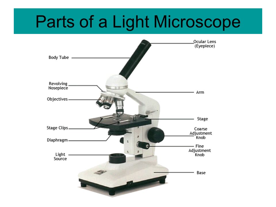
Parts of a Microscope
| Part of the Microscope | Function of the Part |
| Limb | Supports the body tube and stage |
| Base | Provides firm and steady support to the microscope |
| Body Tube | Holds the eyepiece and the revolving nose piece |
| Coarse adjustment knob | Raises or lowers the body tube for longer distances to bring the image into sharper focus |
| Fine adjustment knob | Raises or lowers the body tube through smaller distances to bring the image into sharper focus. It is mostly used with the high power objective lens |
| Diaphragm | An aperture that regulates the amount of light passing through the condenser to illuminate the specimen |
| Eye-piece | Contains a lens which contributes to the magnification of the specimen under review. |
| Objective lens | Brings image into focus and magnifies it |
| Mirror | Reflects light through the condenser to the object on the stage |
| Revolving nose-piece | Holds the objective lenses in place and enables the change from one objective lens to the other |
| Condenser | Concentrates light on the object on the stage |
| Stage | Flat platform where specimen on the slide is placed. It has two clips to hold the slide into position. |
Handling and Care of the Microscope
The following rules should be observed when handling the microscope:
- Always use both hands when carrying the microscope. One hand should hold the base to provide support while the other hand holds the limb.
- Never place the microscope too close to the edge of the working bench or table.
- Do not touch the mirror or the lenses with your fingers.
- Dirty lenses should be cleaned using a special soft lens tissue paper or tissue paper moistened with ethanol. The other parts of the microscope may be cleaned using a microscope.
- Do not wet any part of the microscope.
- Make sure the low power objective lens clicks into position in line with the eye piece before and after use.
- After use, always clean and store the microscope in a safe place, free from moisture and dust.
How to Use the Microscope
- Place the microscope on the bench with the stage facing away from you.
- Turn the low power objective lens until it clicks into position.
- Ensure that the diaphragm is fully open.
- Look through the eye-piece with one eye; meanwhile adjust the mirror under the stage to ensure that maximum light can pass through. The circular area seen is referred to as the field of view.
- Again look through the eyepiece while adjusting the mirror under the stage to ensure that sufficient light is passing through the specimen.
- Use the coarse adjustment knob to bring the low power objective lens to the lowest point. Viewing through the eye-piece, turn the coarse adjustment knob gently until the specimen comes into focus.
- Use the fine adjustment knob to bring the image into sharp focus. Make a drawing of what you observe.
- For higher magnifications, turn the medium power objective lens into position and adjust the focus using the coarse adjustment knob. For sharper images, use the fine adjustment
knob. - If finer details are required, turn the high power objective lens into position; now use only the fine adjustment knob to bring the details into sharper focus.
Cell Structures as Seen under the Light and Electron Microscope
Cell Structure under Light Microscope
- The structures within the cell are referred to as organelles. Some of the cell organelles that can be observed under the light microscope include the cell wall, cell membrane, cytoplasm, nucleus, vacuole and chloroplasts.
- These cell organelles perform specific functions within the cell.
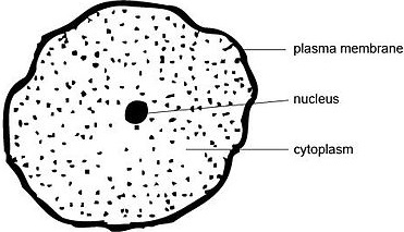
Animal Cell as shown above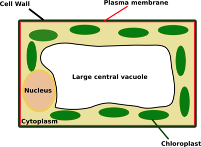
Plant cell as shown above
The Cell as Seen under the Electron Microscope
- The electron microscope is more powerful than the light microscope. It uses a beam of electrons to illuminate the specimen instead of light as in the case of light microscope.
- Electron microscope can magnify an object up to 500,000 times.
- It also has a very high resolving power. Resolving power is the ability to distinguish between separate things which are close to each other.
- The high resolving power makes the electron microscope a very important research tool in microbiology.
- Through the electron microscope, very fine details of the cell can be observed.
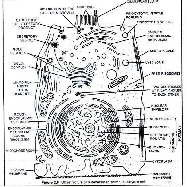
Fig. Animal Cell
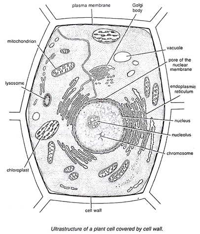
Fig. Plant Cell
Structure and Functions of the Cell Organelles
-
Cell membrane
- The cell membrane, also known as plasma membrane or plasmalemma consists of three layers when viewed under the electron microscope. The three layers are composed of one layer of phospholipid sandwiched between two protein layers.
- It is flexible and has pores. The cell membrane is important in that:
- It encloses the cell contents.
- It allows for selective movement of materials in and out of the cells. The pores allow materials particularly of small molecular size to move in and out of the cells.
-
Cytoplasm
- Cytoplasm consists of a fluid medium in which chemical reactions take place. It contains organelles and other inclusions such as starch, glycogen, fat droplets and many other dissolved substances.
- Cytoplasm is not static; it undergoes a movement known as cytoplasmic streaming.
- It provides a suitable medium for cellular reactions to take place.
-
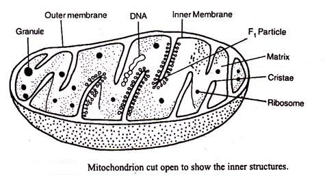
Mitochondrion
- Mitochondrion is a sausage shaped organelle that provides sites for respiratory reactions that yield energy for the cell. Mitochondria is thus, referred to as the powerhouse of the cell.
- It is bound by two membranes. The inner membrane is greatly folded into cristae to increase surface area for respiration.
- The arrangement and number of mitochondria in a cell depends on the cell energy requirements. Cells that require large amounts of energy contain high amount of mitochondria.
- Such cells include muscle cell, sperm cell, apical meristem cells, and kidney cell.
- Mitochondria are self replicative that is they can divide to form new ones.
-
Endoplasmic Reticulum
- Endoplasmic reticulum appears as a series of interconnected channels, running throughout the cytoplasm.
- Their membranes are continuous with the outer membrane of the nuclear membrane.
- Some endoplasmic reticula have granules called ribosomes on their surfaces and are referred to as rough or granular endoplasmic reticula. Endoplasmic reticula that are not associated with ribosomes are called smooth endoplasmic reticula.
- The rough endoplasmic reticulum transports proteins while the smooth endoplasmic reticulum transports lipids.
- Generally, endoplasmic reticula also act as storage areas for synthesized molecules such as enzymes. They also contribute to mechanical support.
-
Ribosomes
- These are spherical in shape. While some are bound to the endoplasmic reticula, some ribosomes are scattered within the cytoplasm (free ribosomes). Their largest dimension is 25 nanometres.
- They are synthesised in the nucleolus.
- They form sites for protein synthesis.
-
Lysosomes
- These are spherical sac-like organelles bound by a single membrane. They contain lytic enzymes which break down large molecules, destroy worn out organelles or even the entire cells.
- Lysosomes also play crucial role in digestion in unicellular organisms.
- The lysosomes are also vital in breakdown of bacteria and other harmful microbes that might have been ingested in food. This explains their high relative abundance in injured or infected cells.
- The membrane of the lysosomes are intact. This is important because if the enzymes leak out, they may destroy the whole cell.
-
Golgi bodies/Golgi apparatus
- These are stacks of membrane bound tube like sacs. They are found close to the cell membrane.
- Golgi bodies perform the following functions:
- They package and transport glycoproteins.
- They are involved in secretion of synthesized proteins and carbohydrates.
- They manufacture lysosomes.
- Note: Golgi bodies are abundant in cells that are active in secretion. For instance pancreatic cells which secrete enzymes and the nerve cells which secrete neurotransmitter substances.
-
Centrioles
- These are rod shaped structures located just outside the nuclear membrane.
- They take part in cell division and also in the formation of cilia and flagella in lower organisms.
- Plant cells lack centrioles.
-
Chloroplasts
- Chloroplasts are egg-shaped structures surrounded by two membranes and contain a gel like stroma through which runs a system of membranes that are stacked together to form grana.
- The granum contains chlorophyll which traps light energy that is used during photosynthesis.
- It is in the chloroplasts that photosynthesis takes place.
-
Vacuoles
- These are sacs that are filled with fluid called cell sap. Vacuoles vary in size.
- Animal cells contain small vacuoles which may be numerous in the cells while plant cells contain one large centrally placed vacuole.
- Sap vacuoles store sugars and salts thereby contributing to the osmotic properties of the cell. This influences how materials move in and out of the cell.
- In some unicellular organisms, food vacuole stores and digests food substances while the contractile vacuole excretes unwanted materials from the cell.
-
Cell wall
- This is the rigid outer cover of plant cells and some lower organisms.
- In plants it is composed of cellulose fibres.
- Cell wall is important in that:
- It gives plant cells their definite shape
- It provides mechanical support and protection against mechanical injury.
- The cell wall allows gases, water and other substances to pass through it.
-
Nucleus
- Nucleus is a double membrane bound structure made up of a viscous fluid known as nucleoplasm in which nucleolus and chromatin materials are suspended. The nuclear membrane has minute pores, nuclear pores which allow materials to move in and out of the nucleus.
- Nucleus controls all the activities of the cell.
- Nucleolus is responsible for manufacture of ribosomes while chromatin contains hereditary materials.
- Nucleus generally takes a sperical or oval shape.
Comparison between Plant Cells and Animal Cells
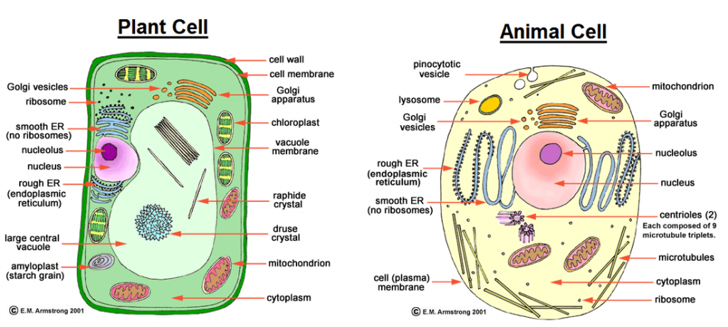
While there exist many similarities between plant and animal cells, there are a number of differences.
| Plant cell | Animal cell |
| Usually large | Smaller in size |
| Regular in shape | Irregular in shape |
| Has a cell wall | Has no cell wall |
| Usually has a large central vacuole | Usually has no vacuoles but when present, they are often temporary and small structures within the cytoplasm |
| Cytoplasm and nucleus are usually located towards the periphery of the cell | Cytoplasm occupies most space in the cell with the nucleus usually centrally placed |
| Some have chloroplasts | Has no chloroplasts |
| Usually more store oils, starch and proteins. | Store glycogen and fats |
| Has no centriole. | Has centrioles |
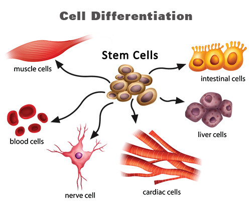
Cell Specialization/Cell Differentiation
- This refers to the process by which a cell becomes structurally modified to perform specific functions
- While cells have a basic outline, they become differentiated to perform specific functions.
- In particular, the root hair cell has extended surface for absorption while the sperm cell has a tail-like extension for swimming towards the ovum.
Tissues
A tissue is a group of cells of a particular type that are grouped together to perform the same function.
-
Tissue types in animals
- Epithelial tissue- This is a thin continuous layer of cells for lining and protection of
internal and external surfaces. - Skeletal muscle - This is a bundle or sheets of elongated cells with fibres that can contract. Its contraction and relaxation brings about movement.
- Blood tissue - This is a fluid containing red blood cells, white blood cells and platelets. The main functions of blood tissue are transportation of nutrients and gases as well as protection of the body against infections.
- Connective tissue - This tissue consists of strong fibres that connects other tissues and organs thereby holding them in position.
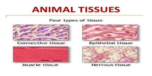
- Epithelial tissue- This is a thin continuous layer of cells for lining and protection of
-
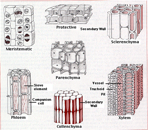
Tissue types in plants
- Epidermal tissue- This is a single thin layer of cells covering the outer surfaces. It protects inner tissues of plants from mechanical damage and infection.
- Palisade tissue- This is a group of cells rich in chloroplasts containing chlorophyll. It has a site for the absorption of light energy and manufacture of food by photosynthesis.
- Parenchyma tissue- This tissue consists of special thin walled irregularly shaped cells. They form packaging and storage cells.
- Conducting tissue/Vascular bundle- This tissue consists of xylem and phloem. Xylem conducts water and dissolved mineral salts in a plant while phloem conducts food substances in solution.
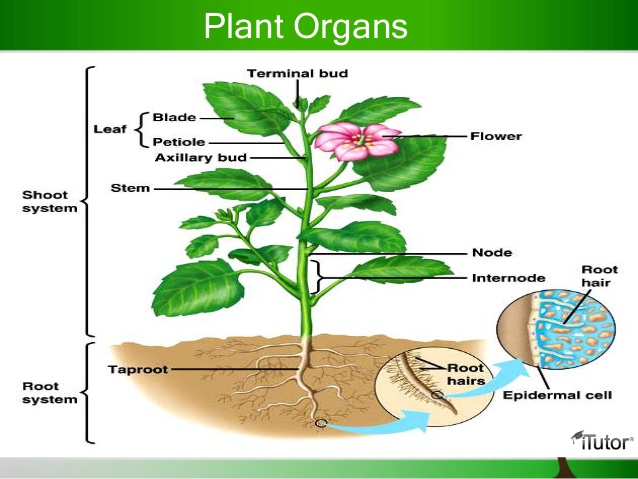
Organs
- An organ is a group of specialized tissues that are grouped together to perform a common function.
-
Organs in animals include:
- Heart- composed of connective, muscle, epithelial and blood tissues.
- Kidney - Composed of connective, epithelial and muscle tissues
- Brain- Composed of epithelial, connective tissues
- Lungs- Composed of epithelial, connective tissues.
-
Organs in plants include:
- Roots- composed of epidermal, conducting and parenchyma tissues.
- Flowers- This is composed of epidermal, conducting tissues.
- Stem- Composed of conducting, parenchyma, and epidermal tissues and palisade tissues in some cases
- Leaves- Composed of palisade, conducting and epidermal tissues.
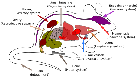
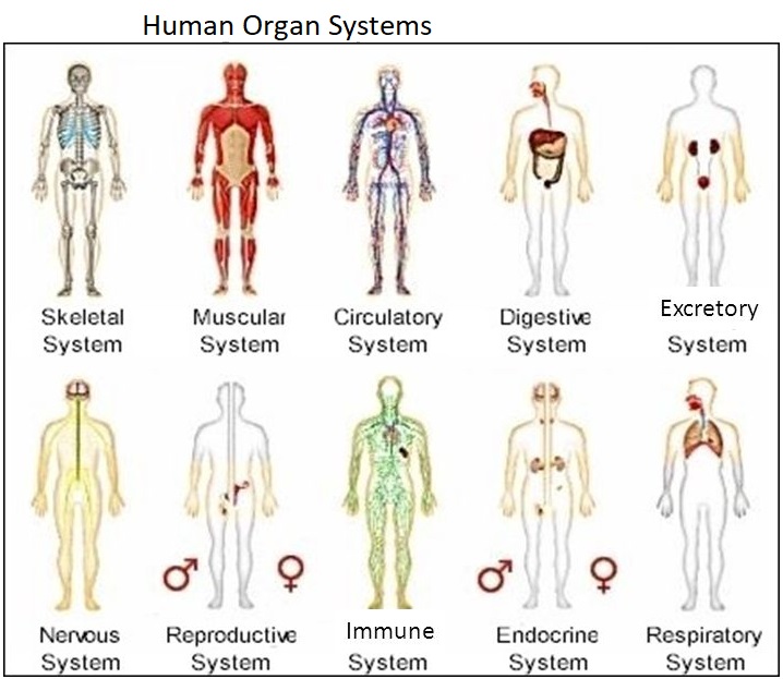
Organ System
- This is a group of organs whose functions are coordinated and synchronized to perform the same function.
- Organ systems are more pronounced in animals than in plants
- Organ systems in animals include
- Digestive system composed of organs such as oesophagus, stomach, intestines and their associated glands.
- Circulatory system composed of the heart, blood vessels (arteries, veins, capillaries).
- Excretory this is composed of kidney, liver, and blood vessels.
- Respiratory system composed of trachea, bronchus, and lungs.
- Reproductive system composed of the reproductive organs and associated glands.
- Nervous systems composed of the brain, spinal cord, eye, ear organs.
Join our whatsapp group for latest updates
Tap Here to Download for 50/-
Get on WhatsApp for 50/-
Download The Cell - Form 1 Biology Notes.
Tap Here to Download for 50/-
Get on WhatsApp for 50/-
Why download?
- ✔ To read offline at any time.
- ✔ To Print at your convenience
- ✔ Share Easily with Friends / Students