
Questions
Instructions To Candidates
- Write your name and index number in the spaces provided at the top of this page.
- Answer all the questions in the spaces provided.
- Study the specimen R provided.
- Identify the type of fruit. (1 mark)
-
- What is the method of dispersal for the specimen R. (1 mark)
- Give reason(s) for your answer in (i) above (2 marks)
- Peel the sliced specimen R to show the inner juicy part. Extract a small portion of the juicy part, place in a mortar and mash it using a pestle.
Filter the extract from the specimen R into a boiling tube.
Divide the extract from specimen R into two portions and use them as follows;
Portion one
Use the reagents provided to test for the food substances present in portion 1. Use the table below as a guide. (6 marks)
Food substance Procedure Observation Conclusion
Portion two -
- To 1cm3 of DCPIP in a test tube, add 0.1% solution of Ascorbic acid drop by drop until the colour of DCPIP disappears. Shake the test tube after addition of each drop. Record the number of drops used. (1 mark)
- To another 1cm3 of DCPIP in a test tube add the portion two of extract drop by drop, shaking the test tube after addition of each drop until the colour of DCPIP disappears. Record the number of drops used (1 mark)
- From the results obtained in (d) (i) and (ii) above, calculate the percentage of Ascorbic acid in the juice obtained from specimen R. Show your working (2 marks)
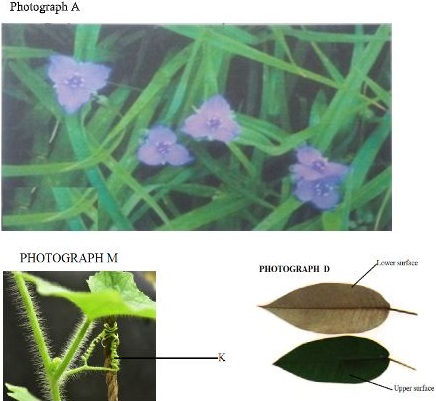
- You are provided with photographs A, M and D representing certain plants and specimens P and Q .Use them to answer the questions that follow.
-
- Name the sub division to which the plant in photograph A and specimen Q belong. (1 mark)
- Give a reason for your answer in a (i) above. (1 mark)
- State the differences between the leaves of specimen P and Q (3 marks)
Specimen P Specimen Q - Name the unique features observed on stems of specimen Q and stem of photograph M and state their function. (2mks)
Specimen Q
Photograph M - Account for the differences observed on the upper and lower surfaces of leaves on photograph D (2mks)
- The stem of specimen Q and that of photograph M are green in colour. What does the colour imply? (1mk)
-
- Name the part labelled K on photograph labelled M (1mk)
- Explain how the coiling of the structure occurred (2mks)
-
-
- Use the photograph provided to answer the questions that follow.
- identify the bone (1mk)
- Give reason for your answer in (i) above (1mk)
- Name the region of the body from which the above bone was obtained (1mk)
- Name the bone which articulates with the above bone at its anterior end (1mk)
- Identify the type of joint formed in (iv) above (1mk)
- Name the structure that joins the two bones in c(i) together at the joint formed above (1mk)
- Identify the view of the above bone in the photograph (1mk)
- State two differences between the above bone and the bone it articulates with at the anterior end. (2mks)
-
- Identify the bone in the photograph below (1mk)
- Name the structure labeled S and state the structure that it articulates with. (2mks)
- Name the structure labelled V and state its function (2mks)
- Name the part labelled M on the diagram (1mk)
- Identify the bone in the photograph below (1mk)
- Use the photograph provided to answer the questions that follow.
Confidential
- Specimen R- a piece of ripe pineapple fruit
- 2mls Benedict’s solution placed in a test tube with a dropper
- 2mls of 10% Sodium hydroxide solution placed in a test tube with a dropper
- 2mls of 1% Copper sulphate solution placed in a test tube with a dropper
- Source of heat
- 4 test tubes in a rack
- 2 Droppers
- Scalpel/Razor blade
- Pestle and mortar
- Filter paper
- A funnel
- A boiling tube
- 4mls DCPIP solution placed in a small beaker with a dropper
- 4mls of 0.1% solution of Ascorbic acid supplied in a test tube
- White tile
- A twig of Lantana camara with flowerslabeled Q
- A twig of Tradescantialabeled P
Marking Scheme
-
- Pome
-
- Animal
-
- Brightly coloured
- Pleasant scent
- Succulent /juicy
- 6mks)
Food substance Procedure Observation Conclusion Reducing sugars Put 2ml of portion 1 into a test tube
Add 2ml of Benedict’s solution
Heat to boilColor changes from blue ,green ,yellow ,orange Reducing sugars present Proteins Put 2ml of portion 1 into a test tube
Add 2ml of Sodium hydroxide solution
Add 3 drops of Copper Sulphateand shake after each dropBlue colour is retained Proteins absent Vitamin C/Ascobic acid To 2ML of DCPIP add food sample drop wise DCPIP decolourised Vitamin C present -
- 4 drops
- Should be more than 4 drops
- 0.1 x 4 drops
drops in d (ii)
- You are provided with photographs A, M and D representing certain plants and specimens P and Q .Use them to answer the questions that follow.
-
- Name the sub division to which the photograph A and specimen Q belong. (1 mark)
- Angiospermaphyta;/acc.Angiospermae Reject if does not begin with capital letter
- Give a reason for your answer in a (i) above. (1 mark)
- They are flower bearing
- Name the sub division to which the photograph A and specimen Q belong. (1 mark)
- State the differences between the leaves of specimen P and Q (3 marks)
Specimen P Specimen Q Network venation Parallel venation Rounded apex Pointed apex Margins are Serrated Margins entire Broader Slender - Name the unique features observed on stems of specimen Q and stem of photograph M and state their function. (2mks)
- Specimen Q
- Presence of spines
- Prevents herbivores from feeding on it.
- Photograph M
- Hairy stem
- Traps a layer of moisture preventing excessive loss of water vapour
- Tendrils
- Support
- Specimen Q
- Account for the differences observed on the upper and lower surfaces of leaves on photograph D (2mks)
- Upper surface is (dark) green while lower surface is white/light grey; chloroplasts are concentrated on the upper surface to trap maximum light for maximum photosynthesis. The light grey lower surface is due to numerous epidermal hairs to reduce transpiration.
- The stem of specimen Q and that of photograph M are green in colour. What does the colour imply? (1mk)
- Have chloroplasts for photosynthesis
-
- Name the part labelled K on photograph labelled M (1mk)
- Tendril
- Explain how the coiling of the structure occurred (2mks)
- Contact on solid object causes lateral diffusion/migration of auxins away from point of contact, higher concentration of auxins at the point away from contact causes faster/rapid cell division and elongation and thus coiling.
- Name the part labelled K on photograph labelled M (1mk)
-
-
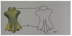
- use the photograph provided to answer the questions that follow.
- identify the bone (1mk)
- Axis
- Give reason for your answer in (i) above (1mk)
- it has odontoid process
- Name the region of the body from which the above bone was obtained (1mk)
- Neck region
- Name the bone which articulates with the above bone at its anterior end(1mk)
- Atlas
- Identify the type of joint formed in (ci) above (1mk)
- Pivot
- Name the structure that joins the two bones in c(i) together at the joint formed above (1mk)
- ligaments
- Identify the view of the above bone in the photograph (1mk)
- Dorsal
- State two differences between the above bone and the bone it articulates with at the anterior end.(2mks)
Atlas Axis Has the articulating facet that articulates with occipital condyles Lacks the articulating facets Lacks odontoid process Has odontoid process
- identify the bone (1mk)
-
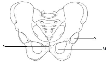
- Identify the bone in the photograph below (1mk)
- Pelvic girdle
- Name the structure labelled S and state the structure that it articulates with. (2mks)acetabulum
- Head of femur
- Name the structure labelled V and state its function (2mks)
- Pubis symphysis
- Name the part labelled M on the diagram (1mk)
- Widens to allow parturition
- Identify the bone in the photograph below (1mk)
- use the photograph provided to answer the questions that follow.
Join our whatsapp group for latest updates
Tap Here to Download for 50/-
Get on WhatsApp for 50/-
Download Biology Paper 3 Questions and Answers with Confidentials - Mokasa II Mock Exams 2022.
Tap Here to Download for 50/-
Get on WhatsApp for 50/-
Why download?
- ✔ To read offline at any time.
- ✔ To Print at your convenience
- ✔ Share Easily with Friends / Students