
BIOLOGY
PAPER 3:PRACTICAL
- You are provided with specimen labelled E and 0.01% DCPIP. Examine specimen E and answer the questions that follow.
-
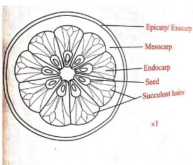
- What part of the plant is the specimen E (1mk)
- Give a reason for your answer in (a)(i) above (1mk)
- Cut a transverse section through specimen E.
- Draw and label one of the cut surface (4mk)
- State the type of placentation of specimen E (1mk)
- State how specimen E is adapted to its mode of dispersal. (2mks)
- Squeeze out the juice from specimen E into a beaker. Filter and discard the residue. Use the reagent provided to test for the food substance present in juice obtained from specimen E. Observe and record in the table below. (3mks)
Procedure observation conclusion
-
- You are provided with two pieces of plant material labeled D. Using a scalpel cut a slip halfway through the middle of each piece as shown in the diagram below.
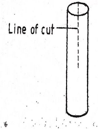
Place one piece in the solution labelled L1 and the other in solution L2. Allow the set up to stand for 30 minutes.- After 30 minutes remove the pieces and press each gently between the fingers
- Record your observations
L1 (1mrk)
L2(1mrk) - Account for the observations in (a)(i) above. (2marks)
- Record your observations
- Examine the pieces.
- Record other observations beside those made in (a) (i)above. (2 marks)
- Account for the observations in (b) (i) above. (6marks)
- After 30 minutes remove the pieces and press each gently between the fingers
- The photograph below represents human male urinary system. Study it carefully and answer the questions that follow.
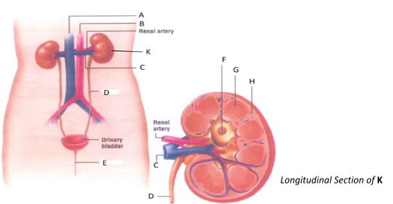
- State two functions of the part labeled K. (2 marks)
- Name the parts labeled A, B, C, D, F, G, and H. (7 marks)
- State the functions of each of the following parts;
- Renal artery (1 mark)
- Urinary bladder (1 mark)
- Part labeled E (1 mark)
-
- State one part of the nephron found in the region labelled G. (1mark)
- Name two kidney disorders (2marks
- Name the hormone that is responsible for reabsorption of water in the renal tubule. (1mark)
CONFIDENTIAL
Each student should be provided with
- An orange labelled as specimen E
- 5Ml 0.01% DCPIP in a test tube
- Scalpel
- Two 50ml beakers
- Dropper
- Sieve
- Two pieces of tradescantia/zebrina stem 3cm long
- A pair of forceps
- 50ml concentrated salt solution labelled L1
- 50ml distilled water labelled L2
- Means of timing
MARKING SCHEME
-
-
- Fruit
- Has two scars
-
-
D=1
L=3
Drawing mark;- continuous outline
-no shading
Labelling marks; -any three correctly labelled parts - Axile placentation
-
-
- brightly coloured
- large and conspicuous
- sweetly scented
- fleshy
-
Note; rej. -marks for observation and conclusion if procedure is wrongProcedure Observation Conclusion Into DCPIP in a test tube add the juice
drop by drop and shakeThe DCPIP is decolourised/blue colour
of DCPIP disappearsAscorbic acid present/vitamin c
-marks for conclusion if observation is wrong
-
-
-
- L1- The piece become less firm/soft OWTTE
L2 The piece becomes more firm/rigid/hard - L1is hypertonic solution cells of the stem loose water through osmosis its cells become flabby/flaccid thus make the stem less firm;
L2 is hypotonic solution cells of the stem gain water through osmosis making the stem more firm;
- L1- The piece become less firm/soft OWTTE
-
- L1 The slit closes and is obscured
L2 Split ends open outward/or curves outward - L1-because the cuticle is water proof;, epidermal cells hardly loose water by osmosis hence their shape does not change;. The cells of the cortex have no cuticle ; hence they lose (a lot of) water by osmosis and shrink;. The differential change in size between the epidermal cells and those of the cortex results in an inward curvature of the split ends obscuring the slit; 4mrks max.3
L2 epidermal cells of the outer surface of the split ends are covered by the cuticle; Hence they take in water by osmosis at a small rate;. Cells of the cortex have no cuticle, hence they take in a lot of water by osmosis and expand;. The differential expansion between epidermal cells and those of the cortex results in an outward curvature of the split ends;.4mrks max.3marks
- L1 The slit closes and is obscured
-
-
-
- excretion
- osmoregulation
- ionic balance
- regulation of pH.
-
- A-Vena cava
- B-Aorta
- C-Renal vein
- D-Ureter
- F-Pelvis
- G-Medulla
- H-Cortex
-
- supply blood to the kidney
- store urine temporarily
- release urine from the urinary bladder
-
- loop of Henle
Collecting tubule - Nephritis
Kidney failure
Kidney stones - Antidiuretic hormone rej. ADH
- loop of Henle
-
Join our whatsapp group for latest updates
Tap Here to Download for 50/-
Get on WhatsApp for 50/-
Download Biology Paper 3 Questions and Answers with Confidentials - MECS Cluster Joint Pre Mock Exams 2021/2022.
Tap Here to Download for 50/-
Get on WhatsApp for 50/-
Why download?
- ✔ To read offline at any time.
- ✔ To Print at your convenience
- ✔ Share Easily with Friends / Students