
Questions
- Which organelle would be abundant in?
- Skeletal muscle cell
- Palisade cell
- State the functions of the following organelles.
- Lysosomes
- Ribosomes
- Name the organelles that perform each of the following functions in a cell.
- Protein synthesis
- Transport cell secretions
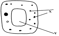
- The diagram below represents a cell.
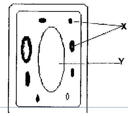
- Name the parts labeled x and y
- Suggest why the structures labeled x would be more on one side than the other side.
-
- State the function of cristae in mitochondria (1mk)
- The diagram below represents a cell organelle
- Name the part labeled Y (1mk)
- State the function of the part labeled X (2 mks)
-
- What is the formula for calculating linear magnification of a specimen when using a hand lens? (1mk)
- Give a reason why staining is necessary when preparing specimens for observation under the microscope. (1mk)
- State three functions of Golgi apparatus. (3mks)
- Name two structures found in plant cell but are absent in animals cell.
- Write the adaptation and role of the following cells.
- Nerve cell
- Palisade cell
- Root hair cell
- Red blood cell
- The diameter field of view of a light microscopic is 3.5mm. Plant cells lying of the diameter are 10. Determine the size of one cell microns (1mm = 1000μm)
- Define the following
- Tissue
- Organ
- Organ system
- Name the organelles that perform each of the following functions:
- Digestion and destruction of worn out organelles.
- Osmoregulation
- Explain why the following processes are important during the preparation of temporary slides
- Staining
- Use of a sharp cutting blade
- In a class experiment to establish the size of an onion cell, a leaner observed the following on the microscope field of view of 4mm. If the student counted 20 cells across the diameter of this field of view, calculate the size of one cell in micrometers.
- State the functions of the following cell organelles:
- Nucleolus.
- Plasma membrane
- What is the of nucleus of a cell made up of?
-
- In a laboratory exercise a student observing a drop of pond water under a microscope saw and drew a spirogyra. If the magnification of the eye-piece was x5 and that of the objective lens was x100, what was the magnification of the spirogyra?
- If the spirogyra has a length of 5cm at the above magnification, calculate the actual length in micrometers
-
- Identify the organelle shown below:-
- How is the organelle you have identified in (a) above suited to its function
- Identify the organelle shown below:-
- Identify the structures of the cells that perform the following functions:-
- Synthesize ribosomes
- Regulate exchange of substances in and out of the nucleus
-
- State the roles of enzyme catalase in living cells
- Which factor inactivates enzyme?
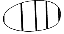
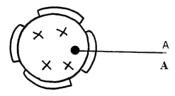
- The figure below represents a certain cell organelle:-
-
- Identify the cell organelle
- What is the function of the part labelled A
- Name the organelles that perform each of the following functions;
- Osmoregulation in amoeba
- Carries out digestion and destruction of worn out cell organelles
-
- State three properties of the cell membrane
- The diagram below represents a plant cell
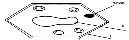
- Name a carbohydrate which forms part of the structure labelled S
- State two functions of the part labelled R
- Name two structures present in the diagram but absent in the animal cell
- What do you understand by the following terms
- Anatomy
- Biochemistry
- State the function of the following parts of a cell
- Ribosome
- Chloroplasts
- What is the formula for calculating linear magnification of a specimen when using a hand lens
- State the function of the following cell structures:-
- Ribosome
- Centrioles ;
- What is the main structural component of:-
- Cell wall
- Cell membrane
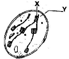
- The diagram below represents a cell
- Name parts labelled X and Y
- Suggest why the structures labelled X would be more on one side than the other
- During a practical class, form fours estimated the field of view to be 3.5mm. Using the low power objective, they observed spirogyra cells across the same field of view and counted 8cells. Calculate the size of each cell and give your answer in micrometer.
- Distinguish between the following terms :-
- Magnification and resolution of a microscope
- Mounting and staining of a specimen
- Name the organelle that performs each of the following functions in a cell.
- Transport of packaged glycoproteins
- Destruction of worn out cell organelles
- Synthesis of proteins
- Why are the following procedures done when preparing sections to be observed under a light microscope?
- Making of thin sections
- Using a sharp blade to make the sections
- Staining
- What are the functions of the following parts of a light microscope?
- Eye piece lens
- Condenser
- Diaphragm
- Given that the diameter of the field of view of a light microscope is 2000um. Calculate the size of a cell in mm if 10 cells occupy the diameter of the field of view
- State the importance of the following processes in microscopy:
- staining
- sectioning
- A cell was found to have the following under a light microscope; cell membrane, irregular in shape, and small vacuoles. Identify the type of the cell above
- State the functions of the following organelles;
- Lysosomes
- Golgi apparatus
- State the functions of each of the following parts in a microscope.
- The eye piece lens
- The objective lens
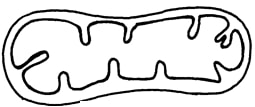
- The figure below represents an electron micrograph of an organelle that is found in many cells;
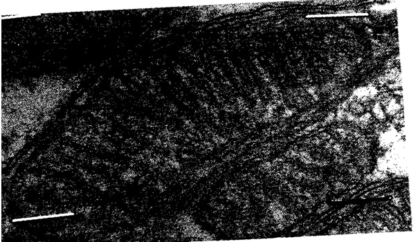
- Identify the organelle
- State the function of the organelle
- What is the importance of infoldings in the inner membrane.
- Give two examples of tissues where you would expect many such organelles in animal body.
Answers
-
- Mitochondrion
- Chloroplast
-
- Destroying old and worn out organelles
- Synthesize proteins
-
- Ribosomes
- Rough endoplasmic reticulum
-
- X- chloroplasts
Y - Vacuole - In dim light. They move to the upper part of the cell in order to receive enough sunlight for photosynthesis
- X- chloroplasts
-
- Increase surface area for attachment of respiratory enzymes hence increasing rate of respiration.
-
- Stroma
- Absorb sunlight used for light stage of photosynthesis
-
- Drawing Magnification = Length of the drawing/Length of the object
- It is adding a dye to the specimen to make the features clearer and distinguishable.
-
- Form vesicles that transport materials to other parts of the cell e.g. proteins
- Transport secretions to the cell surface for secretion e.g. enzymes and mucus.
- They form lysosomes
-
- Cell wall
- Large vacuole
- Chloroplast
- Starch granules
-
- Presence of dendrites to receive impulses
- Presence of chloroplasts to trap sunlight
- Elongated and no cuticles in order to absorb water
- Biconcave shape to increase surface area for diffusion of oxygen/haemoglobin.
- 1 mm = 1000μm
3.5 mm = 3500 μm
10 cells = 3500 μm
1 cell = ?
1 cell = 350 μm -
- Made of several specialized cells grouped together and perform particular function.
- Made of a group of specialized tissues grouped together performing a particular function
- It is made of several organs that perform a particular function.
-
- Lysosomes;
- Contractile vacuoles;
-
- Make cells visible;
- Prevent distortion of cells;
- Diameter of field of view
= 4mm x 1000mm = 4000µm;
Size of each cell = 4000/20
= 200µm; -
- Manufacture of ribosomes;
- encloses cell contents; regulate movement of materials in and out of the cell;
- Protein: Nucleic acid (DNA – RNA);
-
- Mg = O.L.M x E.L.M;
= 100 x 5
= x500; - x 500 = 5 x 10,000 = 50000mµ
x 1 = ?
= (1 x 50,000)/500
= 100micrometer;
- Mg = O.L.M x E.L.M;
-
- mitochondria;
-
- -has cristae/inner membrane highly folded to increase surface area; for respiration.
- -Has matrix medium for respiratory activities; (reject (b) if (a) is wrong.)
-
- nucleolus;
- Centrioles;
- nuclear membrane/pore;
-
- catalyses the breakdown of toxic hydrogen peroxide; to harmless water and oxygen in active tissues;
- Low temperature;
-
-
- Nucleus
- Formation of RNA / ribonucleic acid;
Formation of ribosomes;
-
- Contractile vacuole;
- Lysosomes;
-
- Sensitive to change in temp; sensitive to changes in PH; has both negative and positive charges;
-
- Cellulose;
- Store sugars, salt and food; carry out osmoregulation by inducing osmotic gradient that bring about water movement; maintain the shape of the cell;
- Cell wall; and chloroplast;
- Study of internal and external parts of the body of an organism; Study of the living organisms and their chemical composition;
-
- Synthesis of proteins;
- Site for photosynthesis;
- Length of drawing/Length of object
-
- Ribosomes:- Protein synthesis (1mk);
- Centrioles – Spindle formation during cell division ;
- Form cilia and flagella
-
- cellulose;
- Lipoproteins/lipids and proteins;
-
- X : Chloroplasts; Y : Vacuole /sap vacuole;
- More on the upper side to obtain optimum light intensity/ in bright light, they move away to avoid bleaching/ in dim light they move towards the source of light for maximum absorption of light;
- Cell diameter = field of view in micrometer/Number of cells under the field of view
3.5x1000/8 ; 3500/8;
= 437.6mm = 438mm; -
- Magnification – Ability of a microscope to enlarge tiny objects; Resolution – Ability of a microscope to separate between two tiny structures under magnification to appear distinct
- Mounting – The placing of prepared slide on stage of a microscope; Staining – Use of chemical stain on specimen for clear observation
-
- Golgi bodies/Golgi apparatus;
- Lysosome(s):
- Ribosomes;
-
- Make the sections transparent:
- To produce thin sections/ Not to distort the cells:
- To distinguish between different parts/organelles of the cells:
-
- Magnify the object further;
- Concentrates light onto the object;
- Controls amount of light illuminating the object;
- Size of one cell = diameter of field view/No. of cells arranged across the diameter
= 2000µm/10cells
200µm = 0.2mm
N/B = 1µm = 0.001mms; -
- To make the specimen /section more visible
- To allow light to pass through for easy viewing
- Animal cell;
-
- Stores hydrolytic enzymes for destruction of worn out organelles/ cells/ tissues/ digestion of bacteria/ pathogens; Acc digestion of food/ accept autolysis
- Processing/ packaging synthesized and transporting of packaged cell materials; production of lysosomes/ secretions of packaged material;
-
- magnifying the image of the specimen;
- Objective lens brings the image into focus and magnifies it;
-
- Mitochondria
- early production/ respiration;
- Increases surface area; for attachment of respiratory enzymes;
- Nerve cells; skeletal muscles; cardiac muscles
Join our whatsapp group for latest updates
Tap Here to Download for 50/-
Get on WhatsApp for 50/-
Download The Cell Questions and Answers - Form 1 Biology Topical Revision.
Tap Here to Download for 50/-
Get on WhatsApp for 50/-
Why download?
- ✔ To read offline at any time.
- ✔ To Print at your convenience
- ✔ Share Easily with Friends / Students